
A new study published online in the Journal of Nephrology evaluates the use of acid-free glyoxal in renal ultrastructural diagnostics, highlighting its potential as a safer and more versatile alternative to glutaraldehyde.
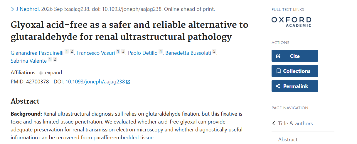
The article, entitled “Glyoxal acid-free as a safer and reliable alternative to glutaraldehyde for renal ultrastructural pathology,” is authored by Gianandrea Pasquinelli, Francesco Vasuri, Paolo Detillo (Lab Manager at Addax Biosciences), Benedetta Bussolati (founder and shareholder of Addax Biosciences), and Sabrina Valente.
Transmission electron microscopy remains an essential tool in the diagnosis of renal diseases, as it enables highly detailed observation of the microscopic structures that make up the glomerular filtration barrier.
The preparation of samples for this type of analysis has traditionally relied on glutaraldehyde fixation. However, this fixative has certain limitations, including a significant toxicity profile and relatively limited tissue penetration.
The study therefore investigated whether acid-free glyoxal could provide adequate preservation of renal ultrastructure and enable diagnostically useful information to be recovered even from samples already embedded in paraffin.
Samples of normal human renal cortical tissue obtained from nephrectomy specimens were fixed in acid-free glyoxal for 24, 48, or 72 hours and subsequently processed for transmission electron microscopy. Samples fixed in 2.5% buffered glutaraldehyde were used as controls.
The researchers assessed the preservation of several key components of the glomerular filtration barrier:
– podocyte foot processes;
– slit diaphragms;
– endothelial fenestrations;
– the glomerular basement membrane.
Samples fixed in acid-free glyoxal, embedded in paraffin, and subsequently recovered and reprocessed for ultrastructural analysis were also examined. Finally, the study assessed the quality of conventional histology, automated immunohistochemistry, and, on an exploratory basis, immunofluorescence.
After 24 hours of fixation, acid-free glyoxal preserved the main ultrastructural components of the glomerular filtration barrier.
Preservation quality improved significantly when fixation was extended to 48 and 72 hours. At these time points, slit diaphragms and endothelial fenestrations were clearly recognizable, with well-balanced image contrast.
A particularly interesting finding concerned tissue recovered from paraffin blocks: these samples also provided adequate ultrastructural detail to address common diagnostic questions. This possibility could offer greater flexibility in sample management, especially when tissue was not originally intended for electron microscopy. Conventional histology was also well preserved.
A Safer and More Versatile Approach
The results indicate that, when appropriate fixation times are used, acid-free glyoxal can provide good to excellent preservation of renal glomerular ultrastructure.
Its ability to yield diagnostically useful information from tissue already embedded in paraffin further strengthens its potential as a versatile fixative for renal pathology.
The study opens new perspectives for introducing safer solutions into ultrastructural diagnostics. It represents another step towards developing fixation processes that combine operator safety, morphological quality, and diagnostic reliability.
A preview of the article is already available on PubMed, pending publication of the final version: